

Cryo-widefield fluorescence of mouse embryonic fibroblast cells on quantifoil gold finder grids [Linkam CLEM workshop at the Crick Institute, London 2015].
Cryo-correlative light and electron microscopy (cryo-CLEM) is a powerful technique that combines the advantages of fluorescence microscopy and electron microscopy to study biological samples at high resolution and in their native state. However, cryo-CLEM poses many challenges, such as reproducible sample preparation, maintaining the sample vitrified and free of contamination, transferring the sample between different microscopes, and locating the same region of interest in both modalities.
Development of the CMS196 & CryoGenium
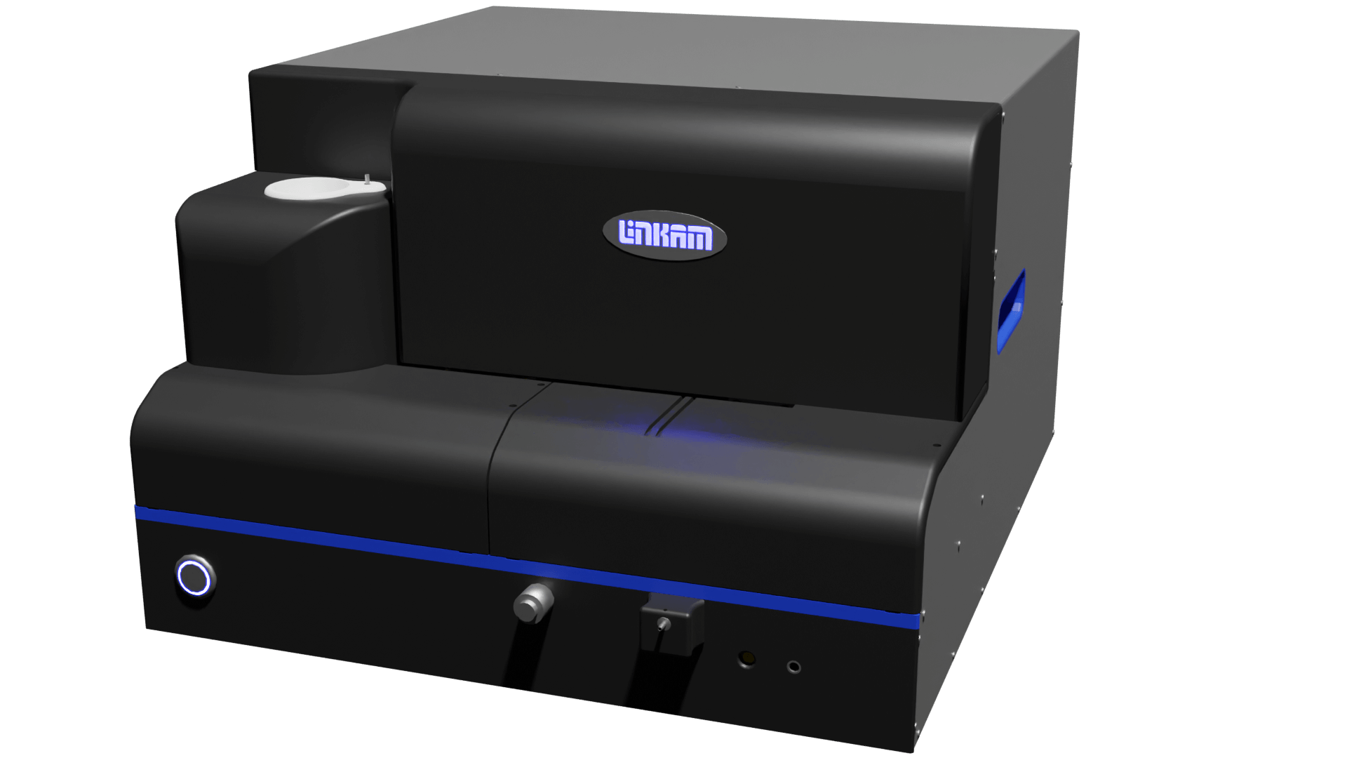
The CMS196 is a cryo-CLEM stage that enables the user to overcome the challenges of obtaining fluorescence microscopy images from the EM grid whilst maintaining the sample at liquid nitrogen temperatures enabling the full workflow of cryo-CLEM. Developed by Linkam Scientific in collaboration with Prof. Bram Koster’s team at Leiden University Medical Centre (LUMC) in the Netherlands, the CMS196 is the result of years of intensive research and development. Linkam is also currently developing the CryoGenium, an automated vitrification robot, to address the issue of inconsistent sample preparation when plunge freezing samples.
CMS196 Applications
In the realm of structural biology, the study of living cells has been revolutionised by the advent of correlative light and electron microscopy. This technique combines the high-resolution structural insights of electron microscopy with the dynamic and specific labelling capabilities of light microscopy. A key aspect of this approach is the use of fluorescent proteins, which allow for the visualisation of specific cellular components in real-time. These proteins can be introduced into living cells, illuminating processes that were previously invisible.
The CMS196 stage has been used for a range of applications in life science research, such as imaging endosomes and autophagosomes in whole mammalian cells, X-ray tomography of cryopreserved human prostate cancer cells [1], multimodal imaging and soft X-ray tomography of fluorescent nanodiamonds in cancer cells [2], imaging of vitrified biological specimens by confocal cryo fluorescence microscopy and cryo FIB/SEM tomography [3], and adaptive 3D cryogenic correlative light imaging of native biology using laser free confocal system [4]. It has also been used to investigate the effect of additives on the effectiveness of cisplatin, a chemotherapy medication, using cryo-fluorescence microscopy [5], and to study intracellular segregation processes in the endocytic system [6]. One of the key benefits of the stage is to pre-screen EM grids using fluorescence microscopy to eliminate damaged grids and to locate regions of interest, reduces wasted time on high demand equipment, such as beamlines [7].
Introducing the CryoGenium
The cryo-plunging techniques most widely used for vitrification are based on paper blotting to adjust the thickness of the vitrified layer. This has been identified as a key issue in the reproducibility of sample preparation because the blotting paper soaks up moisture while inside the high humidity chamber leading to characteristics that are challenging and not always repeatable [8]. Linkam has developed a novel method that overcomes this limitation using an automated plunging robot with real-time optical process control using suction for sample thickness adjustment. The CryoGenium incorporates a glow discharge unit for grid preparation, a dipping station for sample preparation, an optical imaging system with integrating suction for sample thickness control, and a liquid ethane dipping bath for sample vitrification.

Comparison of the traditional paper blotting method of sample preparation with Linkam’s novel suction method
CMS196 Stage

The CMS196 is a self-contained cryo sample observation system that ensures the sample remains vitrified at a liquid nitrogen temperatures. Sample contamination is avoided by the continuous stream of nitrogen gas vaporising from the liquid nitrogen bath surrounding the sample. It can support up to three sample grids of various types, such as EM-grids, Auto-Grids (FEI), Membrane carriers (Leica HPM100), Bessy grids, planchettes, CryoCapsule from CryoCapCell and custom sample holders.
The sample grids are loaded into a specially designed cassette that is transported from a plunge freezer or a high-pressure freezer in a small, sealed container. The container is loaded into a pre-cooled dry sample loading chamber fitted on an upright fluorescent microscope. The cassette is then easily loaded onto the viewing bridge using special manipulation tools.
The CMS196 features an integrated motorised XY stage with optical encoders for high precision movement and position readout of better than 1µm. The stage can be controlled by NEXUS software that enables high resolution maps of the grid to be produced quickly and easily and is also integrated into several microscope manufacturers’ software for automated mapping functionality. For conversion of the co-ordinates in the fluorescence image to EM co-ordinates Linkam provide a simple tool that makes locating regions of interest effortless. This tool is available on our CMS196 webpage.
The CMS196 also has integrated condenser optics for transmitted light brightfield and phase contrast techniques. It is a versatile and efficient instrument that enables routine correlation of fluorescence and EM images. It also optimises optical performance to enable the use of high NA lenses. The CMS196 is compatible with most research grade, upright microscopes from all major manufacturers.
References
[1] Duke, E., et al., “Imaging endosomes and autophagosomes in whole mammalian cells using correlative cryo-fluorescence and cryo-soft X-ray microscopy (cryo-CLXM)”, Ultramicroscopy, 143, 2014, 77-87.
DOI: 10.1016/j.ultramic.2013.10.006
[2] Reineck, P., et al., “Multimodal imaging and soft X-ray tomography of fluorescent nanodiamonds in cancer cells”, Biotechnol. J., 16, 2021, 2000259.
DOI: 10.1002/biot.202000289
[3] Schertel, A., et al., “Imaging of Vitrified Biological Specimens by Confocal Cryo Fluorescence Microscopy and Cryo FIB/SEM Tomography”, EMC 2016, Proc., 2016.
DOI: 10.1002/9783527808465.EMC2016.6998
[4] Nizamudeen, Z., et al., “Adaptive 3D cryogenic correlative light imaging of native biology using laser free confocal system”
Wiley Analytical Science Magazine
[5] Linkam Article: Improved suppression of cancerous cells by regulating the toxicity of chemotherapy medication
[6] Linkam Case Study: Intracellular segregation processes in the endocytic system
[7] Groen, J., et al., “Correlative 3D cryo X-ray imaging reveals intracellular location and effect of designed antifibrotic protein–nanomaterial hybrids”, Chem. Sci., 12, 2021, 15090-15103.
DOI: 10.1039/D1SC04183E
[8] Konig, R.I., et al., “Automated vitrification of cryo-EM samples with controllable sample thickness using suction and real-time optical inspection” Nat. Commun., 13, 2022, 2985.
DOI: 10.1038/s41467-022-30562-7