
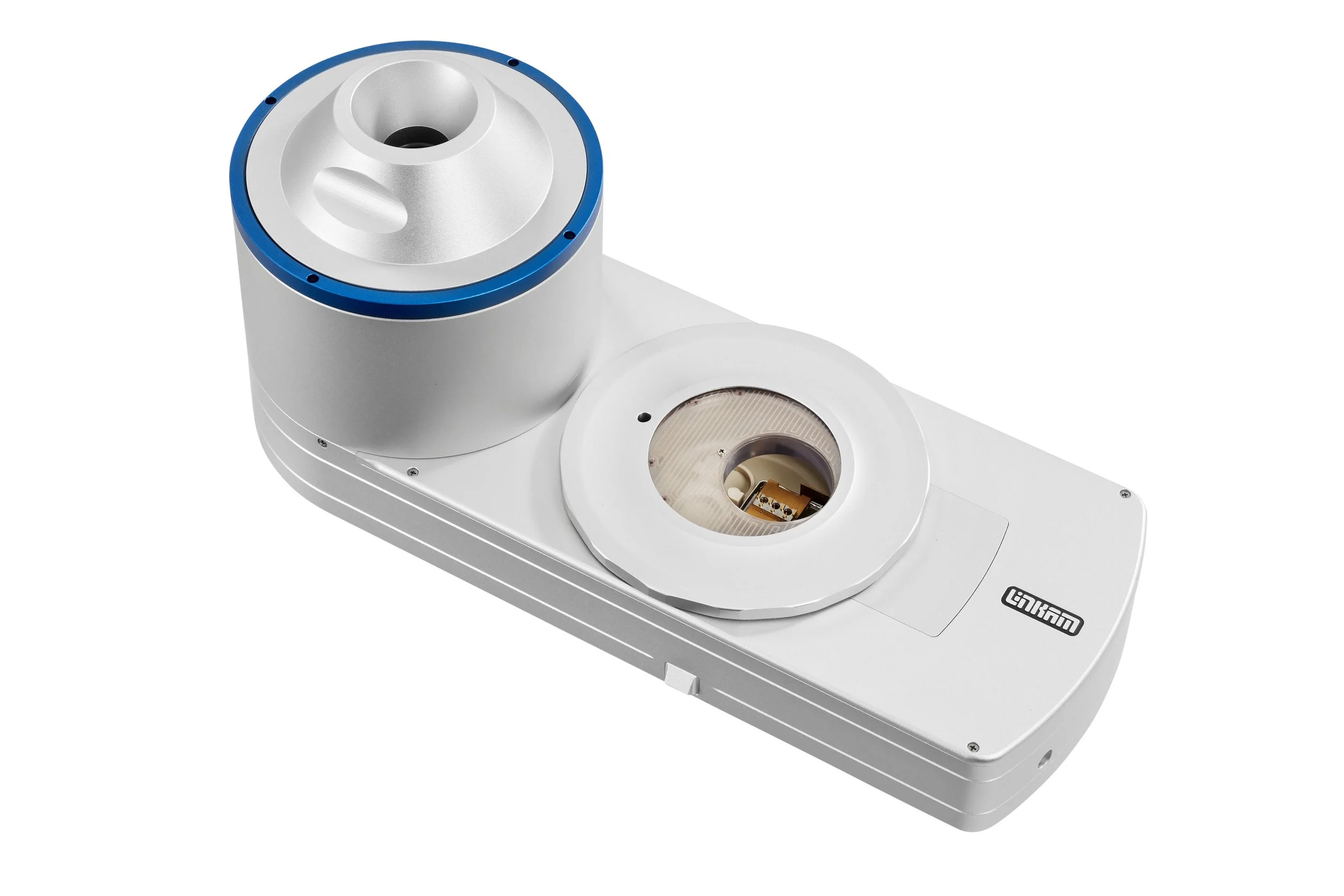
CMS196V4 shown above
Researchers at Hochschule Geisenheim University and the University of Hohenheim in Germany conducted a fascinating study combining cryo-LM and cryo-FIB-SEM techniques to investigate guard cell ultrastructure in higher plants in a near-native state. They developed a workflow that enabled high-resolution 3D visualisation and quantification of chloroplasts, stromules, chloroplast protrusions, mitochondria, vacuoles, and starch granules in Vicia faba guard cells while avoiding the structural distortions often associated with chemical fixation and resin embedding.
A key component of this workflow was a Linkam CMS196 Cryo-Correlative Microscopy Stage used with a Zeiss LSM900 microscope, which allowed cryogenically preserved samples to be imaged under light microscopy at liquid nitrogen temperatures before transfer to FIB-SEM analysis. The CMS196 enabled researchers to assess sample quality, identify regions of interest, visualise chloroplast autofluorescence, and maintain sample integrity throughout the correlative imaging workflow. By preserving samples in a vitrified, near-native state and minimising contamination during transfer, the cryo-stage played a critical role in ensuring accurate ultrastructural analysis.
This study using this near-native volume imaging technique opens new opportunities for studying plant responses to environmental stresses such as drought, salinity, nutrient deficiency, and pathogen attack at the ultrastructural level.
Read the full article here