Linkam stage in Antarctica

Pteropod Limacina helicina antarctica. Photo taken by the Hofmann Group, using a Wild M37 stereomicroscope and Linkam PE120 stage

Pteropod Limacina helicina antarctica. Photo taken by the Hofmann Group, using a Wild M37 stereomicroscope and Linkam PE120 stage

A team from the Hofmann Lab at the University of California, Santa Barbara, are studying the impacts of ocean acidification in calcifying marine invertebrates such as the pteropod Limacina helicina antarctica (pictured).

The team are based at the McCurdo research station in Antarctica, and have been using the Linkam PE120 stage to observe samples both at ambient temperature and at -5°C.

Read more on their research here


Linkam Press Releases October/November 2015

 A post doctorate research associate from Professor Rachel Williams' group at the University of Liverpool uses the Linkam TST350 stage at room temperature to characterise novel polymer materials


A post doctorate research associate from Professor Rachel Williams' group at the University of Liverpool uses the Linkam TST350 stage at room temperature to characterise novel polymer materials

9th October 2015 

Temperature Controlled Stage used in the Characterisation of Novel Electrolytes for more Efficient and less Volatile Fuel Cells

Linkam Scientific Instruments report on the use of their temperature controlled stages in the study of the novel electrolytes that can be used in fuel cells. This work was carried out at the Katholic University of Leuven in Belgium. 

27th October 2015

Tensile Stage Used to Assist with the Development of New Materials that Address Eye Healthcare in the Ageing Population 

Linkam Scientific Instruments report on the use of their tensile temperature controlled stage to develop and characterise novel materials that address the growing burden of eye healthcare in an ageing population. This work is being carried out at the University of Liverpool in the research group of Professor Rachel Williams.

4th November 2015 

Linkam stages in use in the Wolfson Bioimaging Facility at the University of Bristol as part of the endocytic sorting research of Dr Paul Verkade

Linkam Scientific Instruments report on the use of their temperature controlled stages applied to CLEM and fluorescence microscopy to assist in endocytic sorting in the School of Biochemistry at the University of Bristol.


Freeze Drying Training Course

We are pleased to be involved with the Freeze Drying Training Course ‘Lyophilisation: Practical Applications Utilising Latest Equipment’ at the Hooke College of Applied Sciences, running from 3rd-5th November.

This course covers aspects of freeze drying as it relates to the pharmaceutical, biological and food industries. These topics will first be discussed in a classroom setting and then put into practise in an onsite laboratory. Attendees will get hands-on experience with the Linkam FDCS196 freeze-drying cryo-stage.

Details and registration

The Linkam FDCS196 freeze drying system provides the ability to quickly and accurately determine collapse and eutectic temperature and intricately investigate freeze dried structure of complex samples.

Linkam Scientific introduce students to the CMS196 cryo-CLEM stage at the Francis Crick Institute

Towards the end of September we attended the UK’s first Correlative Light and Electron Microscopy (CLEM) interactive workshop. This was held at the prestigious Francis Crick Institute, in London, and will take place in alternate years with the European EMBO CLEM course in Bristol.

The course was fully booked with eight participants from eight different institutes in the UK working in pairs throughout the week to learn different CLEM workflows. The Linkam CMS196 Cryo-Correlative stage was used during the workshop so students could get practical experience of the system and see how it fits into the Cryo-CLEM workflow.

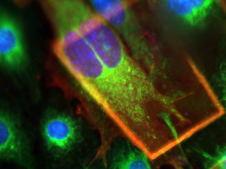
Above is a picture of mouse embryonic fibroblast (MEF) cells, prepared by PhD student Patricia Goggin and captured by Linkam's Michael Schwertner during the ‘correlative cryo-fluorescence leading to cryo-electron tomography or cryo soft x-ray tomography’ practical. This cryofluorescence image shows the cell nuclei in blue, mitochondria in green and filamentous actin in red. 

We had a great time showing off some of the features of our cyro-CLEM stage whilst also continuing to learn about some of its applications, and our thanks go to everyone involved, especially Dr Marie-Charlotte Domart, Dr Raffa Carzaniga, Dr Lucy Collinson and Dr Paul Verkade.