The global outbreak of COVID-19 has prompted scientists all over the world to focus their research on the virus. Some concentrated on understanding the fundamental aspects of the virus and its transmission, while others have focussed on creating vaccines or therapeutics. Many began the search for rapid and robust tests that could be rolled out to a large proportion of the population to diagnose individuals as quickly as possible after infection.
We spoke to Xiaoguang (William) Wang, PhD - recently appointed Assistant Professor in the William G. Lowrie Department of Chemical and Biomolecular Engineering at the Ohio State University (OSU), USA, to discuss his recent ground-breaking work in the field of SARS-CoV2, in particular the development of a rapid detection method for the SARS CoV-2 virus using a liquid crystal (LC) sensor.

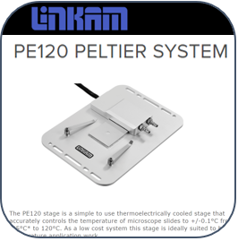
Adil and Yang, members of Prof. Wang’s group at OSU, working with the Linkam PE120 Peltier temperature control setup in the lab.
At the onset of the pandemic, researchers rallied to develop testing protocols for this new and unknown virus. Groups from many different specialties turned their attention to SARS-CoV2 testing, and how they could adapt or pivot their research to tackle this global issue.
Tests using reverse-transcription polymerase chain reaction (RT-PCR) – the ‘gold-standard’ for molecular clinical diagnostics – became available quickly, however, these tests require long characterisation time and specialised equipment to perform. A variety of other technologies have shown promise as candidate diagnostic tools – gold nanoparticles and column agglutination test (CAT), for example – but each has significant limitations. The need arose for fast, simple, and effective techniques to detect and identify the virus.
Professor Wang and the team at OSU responded to the challenge of developing a rapid detection method for the SARS CoV-2 virus using a liquid crystal (LC) sensor. Prof. Wang comments about his decision to consider LCs as a prospective diagnostic technology: “It is commonly known that thermotropic LCs have been employed for the detection of various biomolecules, including DNA and lipids, for example. However, I know of no reports where they have been used for the detection of RNA. So, we set out to apply the technique to SARS-CoV-2 RNA.”
Using thermotropic liquid crystals for viral detection
Liquid Crystals exhibit characteristic and predictable behaviour as a result of their long-range orientational order, and the mobility of their mesogenic constituents. They have been widely utilised in fast-switching electro-optical devices, such as liquid crystal displays (LCDs). They are increasingly used for sensing applications due to their potential for tunable stimuli-responsive properties and self-assembly and ordering characteristics.
Professor Wang’s team at Ohio State university formed a collaboration with Prof. Xiaoping Bao at Purdue University for the design of biosensors for SARS-CoV-2, and Prof. Rongjun Qin at OSU for the development of a smartphone app for automatic readout of the ssRNA of SARS CoV-2.
Summarising the goals of the project, Prof. Wang notes: “LCs are ultrasensitive to small chemical modulations, and different analytes can be detected at extremely low concentrations. Therefore, taking advantage of the high-sensitivity of liquid crystals, we felt that we could develop a rapid detection method for SARS CoV-2 virus that would not need any sophisticated equipment and infrastructure. Moreover, we set out to design a portable and economical LC-based sensor detection kit for the rapid detection of ssRNA of the virus.”
Over the past decade, a series of important works have revealed the design of LC films and droplets that undergo orientational ordering transitions in response to a wide range of molecules adsorbed at an interface. For example, single-stranded DNA (ssDNA) and double-stranded DNA (dsDNA) produce different orientations of LCs at cationic surfactant-laden aqueous-LC interfaces, which leads to a change in the effect on visible light caused by the optical birefringence of the LC film.

Figure 1. Exploded diagram to show the layered make-up of the putative diagnostic device, the LC sensor is show as a dark red spot on the DMOAP glass layer
To show that ssRNA from the virus exhibited the same property, and that LC’s were suitable for COVID detection, Prof Wang’s group followed a 4-step experimental programme.
Step 1. A cationic surfactant dodecyltrimethylammonium bromide (DTAB)-decorated interface on micrometer-thick films of a commercially available nematic liquid crystal, “E7”, was prepared. The layers were supported on a dimethyloctadecyl[3-(trimethoxysilyl) propyl]ammonium chloride (DMOAP)-functionalized glass slide, which induced a perpendicular ordering of the E7.
Step 2. A ssDNA probe, with a sequence complimentary to the defined SARS-CoV-2 ssRNA target, was adsorbed at the DTAB-decorated aqueous-E7 interface. The negatively charged ssDNA was attracted to the cationic DTAB at the aqueous-E7 interface via electrostatic interactions.
Step 3. ssRNACoV was added to the DTAB-laden E7 surface with the adsorbed ssDNAprobe. The temperature of the system was increased to 48.7°C, which is the Tm of the ssRNACoV. A Linkam PE120 Peltier hot stage was used to control the temperature of the E7 surface during these measurements.
Step 4. The optical appearance (i.e., brightness) of the RNA-adsorbed E7 films was quantified from images captured by a CCD camera on a polarized microscope.
Prof Wang summed up the success of these initial experiments, saying: “Our work has shown that LC-based sensors offer a highly sensitive, reproducible and robust method of detection of target ssRNA cov – and we could establish the detection limit of the E7 surface for the target ssRNA by varying the concentration from nanomolar to femtomolar amounts.”
Figure 2 shows a schematic illustration of the optical response of the DTAB/ssDNAprobe-decorated LC film to the adsorption of ssRNAcov.

Figure 2. Schematic showing the physical arrangement of LCs – left shows alignment pre-addition of ssRNAcov; right shows the change in alignment induced by the binding of the ssRNAcov target to the ssDNA probe. Note: the insert image captures show the visual change from occluded (left) to clear (right).
Prof. Wang characterised the Linkam PE120 stage as providing many advantages in their work to detect the ssRNAcov target: “The PE120 gave us not only the precise and reproducible temperature control we needed, but it was also very straightforward to integrate with our microscope and other equipment. The touch display on the controller made it easy and smooth to work with. Moreover, the Linkam stage is highly portable and can be transferred from one lab to another.”
Building a sensor – smartphone detection of the target ssRNA
With the principle of sensor structure and detection performance established, the group moved on to prepare a final sensor design (Fig. 2) that included polarising layers to enable detection of the confirmation change in the LCs by visible light. In addition, the sensor device was integrated with a retaining clip and an appropriate smartphone app to ensure consistent illumination of the sensor. This ensures that non-expert users can reliably read and act upon the results of a test (Fig. 4).

Figure 4. A smartphone screenshot indicating a negative result (left) and positive result (right)
The immediate next step for the COVID work is to move the test into a clinical evaluation phase – testing real patient samples in a biosafety level 3 (BSL-3) laboratory.
However, due to limited access to the labs throughout the pandemic, the group has been unable to perform those studies to date. Assuming satisfactory results of this next stage, and completion of the validation of the system in its practical application, Prof. Wang hopes to extend the collaboration to include the diagnostics industry for further studies and commercialisation, commenting: “We are optimistic that when the current phase of the pandemic is over and work in the labs is resumed normally, an LC-based test could be in the market in the relatively near future.”
In conclusion, when asked about other fields where this methodology could be used, Prof. Wang added: “I believe that this prototype of ‘LC sensor combined with smartphone app’ can be used for the detection of a wide variety of biomolecules and toxic compounds in the near future.”
The detailed experimental protocols, results and a discussion of the significance of this work has recently been published here.
Xu, Yang, et al. "Ultrasensitive and Selective Detection of SARS-CoV-2 Using Thermotropic Liquid Crystals and Image-Based Machine Learning." Cell Reports Physical Science (2020): 100276.
This article will be published in full in Biophotonics.
Related Content