A recent study undertaken at Rovira i Virgili University in Tarragona, Spain, reveals that biolabeling through luminescence thermometry has the potential to detect cancer cells at an early stage of development by monitoring the localised temperature rise in the body, due to accelerated metabolic activities in abnormal cells.
The researchers synthesised core-shell nanoparticles that display temperature-dependent fluorescence. By characterising the change in emission spectra using UV-VIS-NIR spectroscopy with the THMS600, they are able pinpoint minute temperature changes at the cellular scale, bringing the possibilty of using these nanoparticles as in-vitro nanothermometers inside cells.
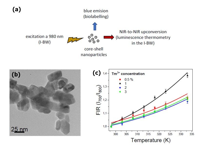
Schematic representation of the pumping and emission bands generated by the Tm,Yb:GdVO₄@SiO₂core-shell nanoparticles, to illustrate their practical use as biolabels, with emission in the blue, and luminescent thermometers operating in the first biological window (I-BW). (b): Transmission electron microscope (TEM) image of the annealed Tm,Yb:GdVO₄ nanoparticles (NPs). (c): Normalized fluorescence intensity ratio (FIR) as a function of temperature for the Tm,Yb:GdVO₄@SiO₂ core-shell nanoparticles doped with different concentration of Tm³⁺. Image used from ref 1 under CC BY 4.0.
The researchers used the Fluorescence Intensity Ratio (FIR) technique to characterise the relationship between the temperature and emission spectra of Tm³⁺ doped SiO₂ core-shell nanoparticles. They excited the particles with a near-infrared laser while using the THMS600 to control the particle temperature, cycling between 298 and 310 K, while observing the resulting emission spectra of the fluorescing particles in the visible region, which allowed them to reproducibly and precisely characterise the FIR as a function of temperature. During testing, the sample was placed on top of a boron nitride disc to aid the uniform and fast distribution of temperature to ensure that the entire sample was maintained at the same heat. Temperature stability and control is a critical aspect in the calibration of the development of the luminescent nanothermometers. The well-characterised highly thermally sensistive particles are also biologically compatible due to the inert silica outer shell surrounding the fluroescent inner core.
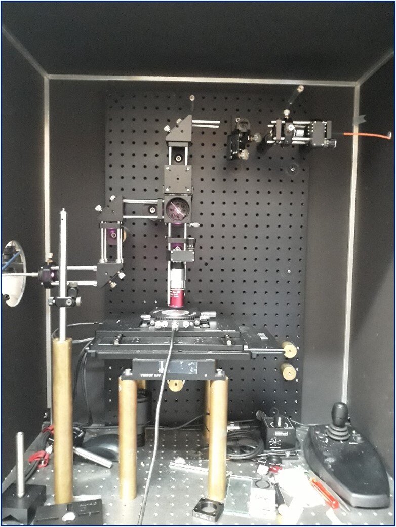
The temperature controlled imaging setup in the Faculty of Chemistry at Rovira i Virgili University
Joan Josep Carvajal Marti, Vice Dean of the Faculty of Chemistry at Rovira i Virgili University comments: “The use of the stage allowed us, among other things, to develop luminescent thermometers operating in the different optical biological windows located in the near infrared part of the electromagnetic spectrum. This is where biological tissues are more transparent to the light, attaining deeper penetration depths which would allow the future treatment of diseases, such as cancer, through laparoscopic techniques.
“We have also been able to develop thermochromic thermometers, in which the colour of the emission changes in accordance with temperature, and that can be used as naked eye indicators to demonstrate if industrial processes are progressing correctly or not.”
Temperature control stages are well suited for the analysis of temperature-sensitive materials such as these luminescent particles. They can be used in a range of applications, from biological samples to photovoltaics, where precise temperature accuracy and stability are required as well as the ability to image the sample through microscopy or analyse using spectroscopic techniques. This is the case for the research undertaken at Rovira i Virgili University where a wide temperature range and pinpoint accuracy were necessary. This work provides essential insight into the behaviour of temperature-responsive particles for cancer detection, and we are pleased to help enable such research.
The techniques using the THMS600 developed by the research team at Rovira i Virgili University enable, for the first time, the measurement of the thermal resistance of a nanoparticle. This is a milestone that opens up research into the thermal properties of nanomaterials, allowing researchers to establish strategies to tailor these properties through the suitable design of the materials. The scientific breakthroughs by this group are making a vital contribution to the advancement of tools to detect, understand and treat cancer.
References
“Bifunctional Tm³⁺,Yb³⁺:GdVO₄@SiO₂ Core-Shell Nanoparticles in HeLa Cells: Upconversion Luminescence Nanothermometry in the First Biological Window and Biolabelling in the Visible”. Oleksandr Savchuk,Joan Josep Carvajal Marti, Concepción Cascales, Patricia Haro-Gonzalez, Francisco Sanz-Rodríguez, Magdalena Aguilo and Francesc Diaz Nanomaterials 2020, 10(5), 993 https://doi.org/10.3390/nano10050993
Related Content