CMS196 application NOTES
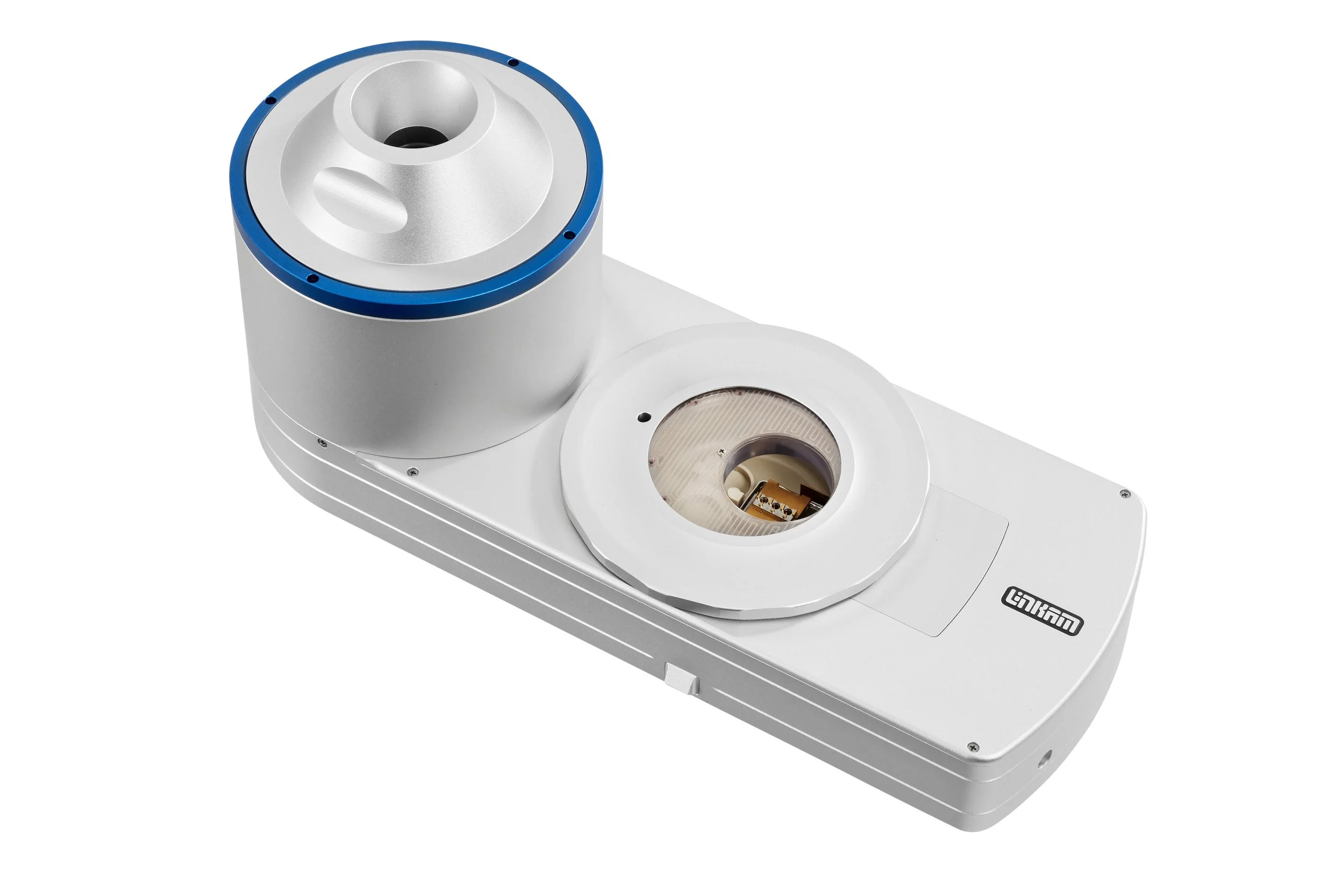
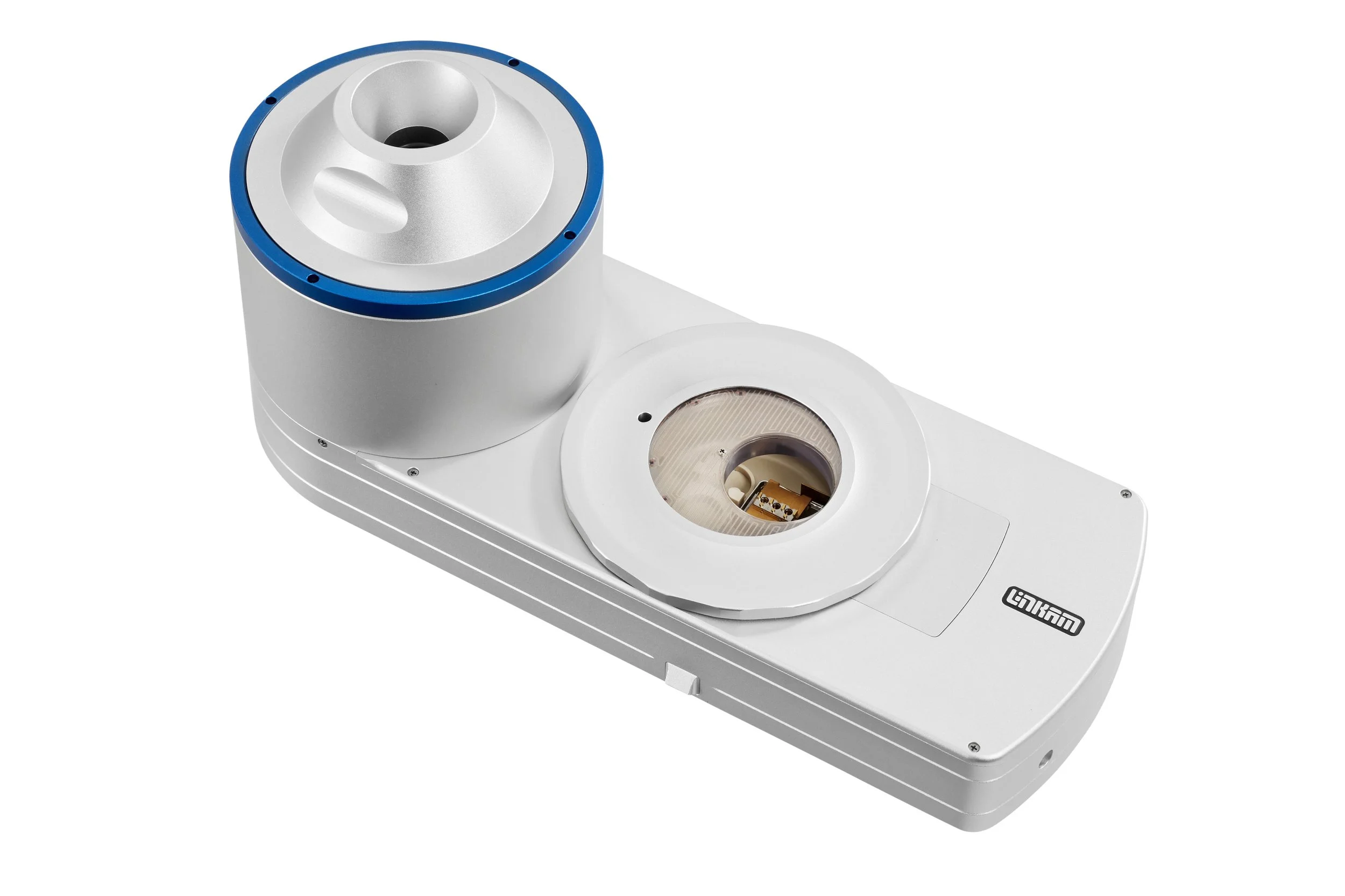
Researchers at King’s College London in the UK supported the development of early models of our CMS196 V4 stage and continue to test new cryomicroscopy systems including the CryoGenium.
Researchers led by Dr Maria Harkiolaki at Diamond Light Source in the UK investigate some of the world’s most challenging biological issues using Linkam’s state of the art sample stages.
Linkam stages including the CMS196 in use in the Wolfson Bioimaging Facility at the University of Bristol as part of the endocytic sorting research of Dr Paul Verkade.
Researchers in Barcelona investigate the effect of additives on the effectiveness of cisplatin, a chemotherapy medication, using the CMS196 for cryo-fluorescence miscroscopy.
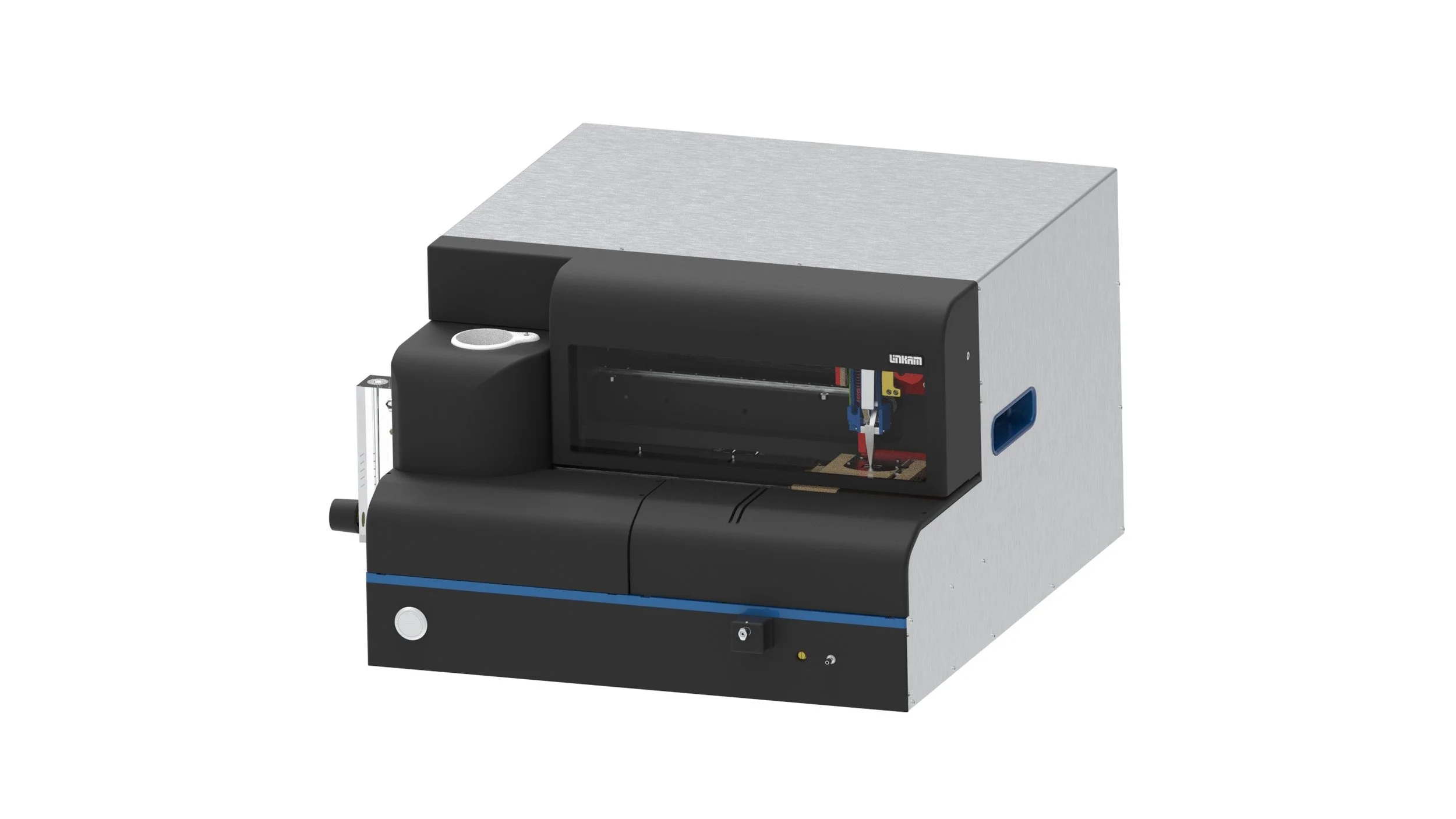
Preparation and handling of vitrified samples normally requires special skills and techniques. Here, we discuss a novel design that simplifies and automates plunge-freezing and sample transfer, thus making this technique accessible for a wider audience.
CMS196 Research Papers
Groen et al. explored how a novel 3D cryo-correlative light and X-ray tomography (CLXT) imaging approach could be used to investigate a promising antifibrotic treatment for myocardial fibrosis, using Linkam’s CMS196 to pre-screen vitrified grids.
Using a Linkam CMS196 stage Beer et al. demonstrated an innovative, targeted cryoCLEM workflow for tissues, in which cryogenic confocal fluorescence imaging of millimetre-scale volumes is correlated to 3D cryogenic electron imaging directed by a patterned surface generated during high-pressure freezing (HPF).
Cryogenic light microscopy is a key mapping step in the correlative imaging workflow. Researchers adapted a standard upright widefield microscope into a cryogenic 3D laser-free confocal system using Linkam’s CMS196, to demonstrate the necessary sample preparation steps followed by confocal imaging of biological cells and tissues.
Researchers used the Linkam CMS196 for imaging cryopreserved human prostate cancer cells with X-ray tomography.
Scientists at the The Institute of Cancer Research in London, UK used Linkam’s CMS196 to image mammalian cells with cryo-CLXM microscopy.
Researchers used Linkam’s CMS196 as part of a Confocal Cryo Fluorescence Microscopy setup to image vitrified biological samples.


