
Cardiac fibrosis is a major health concern that affects millions of people worldwide and has a significant impact on the progression of many heart diseases. Despite its prevalence, there is currently no preventive or curative treatment for this condition due to the elusive mechanism of fibrosis and the lack of specific targets. However, recent developments in imaging technology and therapeutic agents offer new hope in the fight against cardiac fibrosis. In their recent paper Groen et al. explored how a novel 3D cryo correlative light and X-ray tomography (CLXT) imaging approach could be used to investigate a promising antifibrotic treatment for myocardial fibrosis, using a Linkam CMS196 to pre-screen vitrified grids. By incorporating this stage into their workflow, the researchers were able to remove damaged grids and locate cells of interest, ultimately saving valuable beamline time and increasing their efficiency.
Precise targeting for 3D cryo-correlative light and electron microscopy volume imaging of tissues using a FinderTOP
3D high-resolution imaging is essential for understanding the structural organisation and functioning of cells and tissues. Cryo-correlative light and electron microscopy (cryoCLEM) is an approach that combines fluorescence microscopy with cryo-electron tomography (cryoET) to resolve the structure of proteins within their native cellular environment. This approach is mostly used for 2D cell cultures, but there is a growing demand for 3D biological model systems, such as organoids. High-pressure freezing (HPF) can be used to vitrify samples up to 200 μm in thickness, but the featureless ice surface of HPF tissue samples makes it difficult to precisely correlate light and electron images. CryoFIB/SEM volume imaging allows for detailed nanoscale investigation of vitrified samples with multi-micron dimensions, but cryoCLEM volume imaging of tissues is still to be explored. Cryogenic fixation and imaging can overcome some of the drawbacks of room temperature CLEM volume imaging, allowing for the visualisation of cells and tissues in their native state in their natural environment.
Using a Linkam CMS196 stage Beer et al. have demonstrated an innovative, targeted cryoCLEM workflow for tissues, in which cryogenic confocal fluorescence imaging of millimetre-scale volumes is correlated to 3D cryogenic electron imaging directed by a patterned surface generated during high-pressure freezing (HPF). They applied this workflow to study the mineralisation process in scales of zebrafish as a model system for 3D organs. The scales form an interesting model to study bone formation processes, as the elasmoblasts remain active and vital for hours after being removed from the skin. This workflow allowed for uncompromised imaging of tissues in their near-native state over all relevant length scales, from the millimetre down to the nanometre level, opening up future avenues to study structure-function relations of biological materials, in health and disease.

Automated vitrification of cryo-EM samples with controllable sample thickness using suction and real-time optical inspection
Imaging of biological samples embedded in vitrified ice has become of great interest in recent years as it provides several advantages: the biological sample is in a fully hydrated state with superior preservation down to ultra-structural level, a vitrified sample is naturally compatible with the vacuum required for EM / Single Particle (SPT) / CLEM (Correlative Light and Electron Microscopy) and cryo-fluorescence provides very low photo-bleaching and high signal to noise imaging.
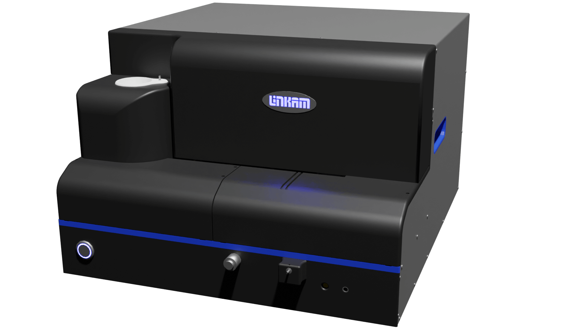
Preparation and handling of vitrified samples normally requires special skills and techniques. The novel design of the Linkam CryoGenium makes this a simple and reproducible process.
Small Molecule Ice Recrystallization Inhibitors Enable Freezing of Human Red Blood Cells with Reduced Glycerol Concentrations

Intrinsic mechanical behavior of femoral cortical bone in young, osteoporotic and bisphosphonate-treated individuals in low- and high energy fracture conditions

A key function for microtubule-associated-protein 6 in activity-dependent stabilisation of actin filaments in dendritic spines

X‐ray diffraction studies of the structural organisation of prolamellar bodies isolated from Zea mays

Human bone marrow stromal cell responses on electrospun silk fibroin mats

The Kinetics of Thermal Injury in Human Renal Carcinoma Cells

Intracellular ice formation in human oocytes vs. cooling rate and temperature

Febrile Temperature Elevates the Expression of Phosphatidylserine on Plasmodium falciparum (FCR3CSA) Infected Red Blood Cell Surface Leading to Increased Cytoadhesion

Adaptive 3D cryogenic correlative light imaging of native biology using laser free confocal system
Cryogenic light microscopy is an important part a correlative imaging workflow, allowing confirmation of localised molecules of interest prior to processing for downstream analysis such ultrastructural or compositional assessment or in short: It is a key mapping step.
Here, the authors describe adaptation of a standard upright widefield microscope into a cryogenic 3D laser-free confocal system using the Linkam CMS196. We demonstrate the necessary sample preparation steps followed by confocal imaging of biological cells and tissues.
Access the full version here: https://analyticalscience.wiley.com/do/10.1002/was.00170086
Nizamudeen, Zubair Ahmed, et al. "Adaptive 3D cryogenic correlative light imaging of native biology using laser free confocal system. Wiley Analytical Science

An Image is Everything: A Tutorial on Choosing and Using the Components of a Dynamic Data Capture System
Linkam provide sample characterisation solutions across a range of temperature and environmental control methods which can be used in combination with microscopic and spectroscopic analysis techniques. Here, we share our expertise in temperature-controlled microscopy with a discussion of how to assemble a dynamic data capture and imaging system.
Access the full version here: https://doi.org/10.1017/S1551929520001558
Stacey, D., & Gurney, R. (2020). An Image is Everything: A Tutorial on Choosing and Using the Components of a Dynamic Data Capture System. Microscopy Today, 28(6), 44-49. doi:10.1017/S1551929520001558

Intrinsic mechanical behavior of femoral cortical bone in young, osteoporotic and bisphosphonatetreated individuals in low- and high energy fracture conditions
Researchers use Linkam’s mechanical testing stage to investigate treatments for osteoporosis in fractures of femoral bones.
Full Open Access article availble via CC BY
Zimmermann, E., Schaible, E., Gludovatz, B. et al. Intrinsic mechanical behavior of femoral cortical bone in young, osteoporotic and bisphosphonate-treated individuals in low- and high energy fracture conditions. Sci Rep 6, 21072 (2016). https://doi.org/10.1038/srep21072

Multimodal Imaging and Soft X‐Ray Tomography of Fluorescent Nanodiamonds in Cancer Cells
Linkam’s CMS196 is used in many biological experimental procedures, such as this work from a group at RMIT University in Melbourne, Australia where the CMS196 is used to study the cell morphology of fluorescent nanodiamonds (FNDs) with confocal fluorescence microscopy. They observe uptake of these FNDS by cancer cells, opening up the possibility of their use in biological imaging and sensing.
Read the full article here (access required).

X-ray tomography of cryopreserved human prostate cancer cells: mitochondrial targeting by an organoiridium photosensitiser
Researchers use the Linkam CMS196 for imaging cryopreserved human prostate cancer cells with X-ray tomography. The CMS196 was used for cryo-fluorescence imaging of human PC3 prostate cancer cells, bringing insight into a new material used for a promising new non-invasive theraputive cancer treatment technique, Photo-Dynamic Therapy (PDT).
Full article available per CC BY 4.0.
Bolitho, E.M., Sanchez-Cano, C., Huang, H. et al. X-ray tomography of cryopreserved human prostate cancer cells: mitochondrial targeting by an organoiridium photosensitiser. J Biol Inorg Chem 25, 295–303 (2020). https://doi.org/10.1007/s00775-020-01761-8

Imaging endosomes and autophagosomes in whole mammalian cells using correlative cryo-fluorescence and cryo-soft X-ray microscopy (cryo-CLXM)
Researchers use the Linkam CMS196 with correlative cryo-fluorescence and cryo-soft X-ray microscopy (cryo-CLXM) to observe endosomes and autophagosomes in mammalian cells.
Scientists at the London Research Institute of Cancer Research UK use the Linkam CMS196 for imaging mammalian cells with cryo-CLXM microscopy.

Imaging of Vitrified Biological Specimens by Confocal Cryo Fluorescence Microscopy and Cryo FIB /SEM Tomography
Researchers use the Linkam CMS196 as part of a Confocal Cryo Fluorescence Microscopy setup to image vitrified biological samples.
Schertel, Andreas, et al. "Imaging of Vitrified Biological Specimens by Confocal Cryo Fluorescence Microscopy and Cryo FIB/SEM Tomography." European Microscopy Congress 2016: Proceedings. Weinheim, Germany: Wiley‐VCH Verlag GmbH & Co. KGaA, 2016.

Effect of supercooling and cell volume on intracellular ice formation
Studying the effect of supercooling and cell volume on intracellular ice formation (IIF), using Linkam’s Freeze Drying system FDCS196.
Full article available per CC BY 4.0.
Prickett, Richelle C., et al. "Effect of supercooling and cell volume on intracellular ice formation." Cryobiology 70.2 (2015): 156-163.

Small Molecule Ice Recrystallization Inhibitors Enable Freezing of Human Red Blood Cells with Reduced Glycerol Concentrations
Researchers use Linkam’s Freeze Drying system FDCS196 to cryopreserve red blood cells with slow cooling rates prior to storage at -80°C.
Full article available per CC BY 4.0.
Capicciotti, C.J. et al. Small Molecule Ice Recrystallization Inhibitors Enable Freezing of Human Red Blood Cells with Reduced Glycerol Concentrations. Sci. Rep. 5, 9692; DOI:10.1038/srep09692 (2015).
