Optical profilometry is a rapid, non-destructive, and non-contact surface metrology technique, which is used to establish the surface morphology, step heights and surface roughness of materials. It has a wide range of applications across many fields of research, including analysing the surface texture of paints and coatings, analysing micro-cracks and scratches and creating wear profiles for structured materials including micro-electronics, and characterisation of textured or embossed nanometre-scale semiconducting components, such as silicon wafers.
Historically, it has been difficult to conduct temperature-controlled optical profilometry experiments due to imaging issues caused by changes in spherical aberration with temperature of both the front lens of the objective and the quartz window of the LTS420 stage.
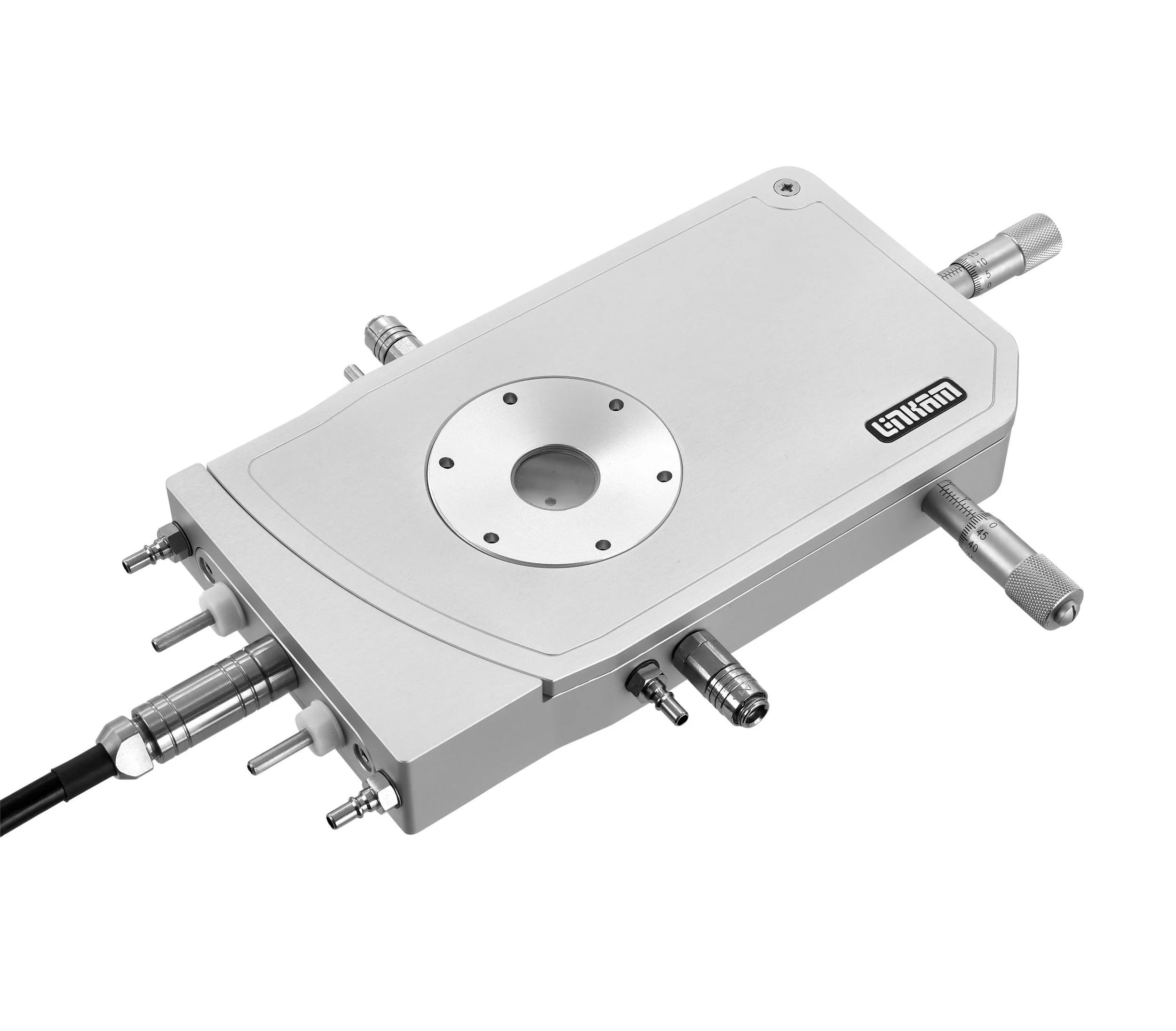
To provide a solution for temperature-controlled optical profilometry Linkam has partnered with Sensofar, who specialise in the field of non-contact surface metrology, to develop a new technique for characterising the evolution of a sample’s surface topography with temperature using the S neox 3D optical profiler and Linnik interferometer coupled with Linkam’s LTS420 temperature-controlled chamber. The technique has been used to successfully map the changes in roughness and waviness of silicon wafers at temperatures up to 380°C.
Linkam LTS420 and Sensofar Linnik configuration
By using Sensofar’s new Linnik interferometer lens system with the S neox 3D optical profiler, in combination with Linkam’s LTS420 precision temperature control chamber, spherical aberration issues are resolved, enabling the accurate measurement of 3D topographic profiles of nanoscale materials at a wide range of temperatures.
For the design and construction of the Linnik objective, two Nikon 10x EPI objectives (Nikon, MUE12100) with 17.5mm working distance were used. The same configuration is available with 10xSLWD objectives (Nikon, MUE31100), providing a 37mm working distance.
This makes the thermal emissions from the camera almost imperceptible to the lens and will not affect or damage the measurement quality. The Linnik objective was mounted on the 3D optical profilometer (Sensofar, S neox), which combines 4 optical technologies in the same sensor head: Confocal, CSI, PSI and focus variation. These techniques are covered in ISO25178.
The example below shows how the 1D profilometric data can be plotted in the form of a topographic image. By stacking the 3D images as a function of temperature, it is possible to create a “4D plot”, as shown in the stacked image below.
This shows the evolution of the topographical changes at different temperatures using a colour scale to indicate height in the vertical direction. In the silicon wafers tested, it is clear that the samples bend as temperature changes. As temperature increases above room temperature towards 380°C, greater bending is experienced by the samples.
Bow evolution in (a) sample A and (b) sample B as a function of temperature. Waviness parameters Wz were extracted from the horizontal, diagonal and vertical profiles in Figure 5. Roughness parameter Sz was computed from the surface after applying an S-filter of 0.8 mm.
Stacked 4D view of the topographies extracted from (a) sample A and (b) sample B for visual comparison of the experimented bow change when samples increase from 30ºC to 380ºC.
The feasibility of the proposed configuration has been proven to carry out successful roughness and waviness measurements at different temperatures. Two different behaviours of the surface topography were observed depending on the chip design. Sample A showed an early bending behaviour when heating up the sample, whereas sample B showed the bending in a later stage.
David Páez, Sensofar Sales Support Specialist, commented: “In a recent experiment using the new technique, we were able to observe the changes in topography of silicon wafers as they evolve with temperature from 20°C up to 380°C. This is critical information for silicon wafer producers and users, so that they can optimise their process, improve semiconductor properties and wafer durability. The Linkam LTS420 chamber and T96 temperature controller are key components in our experimental set-up and enable us to ramp and control the temperature between <-195° and 420°C to a precision of 0.01°C.”
Linkam systems have provided precise temperature and environmental control to a wide range of techniques, from microscopy to X-ray analysis, for decades. This collaboration with Sensofar highlights the important role of temperature control in contributing to innovative approaches to material characterisation.
We are delighted to be able to offer a solution for temperature-controlled profilometry using Sensofar’s Linnik interferometer, and we look forward to seeing how this new technique helps scientists across many fields to advance their research.
Using the Sensofar Linnik system with the LTS420
Related Content


